(C) 2010 Lucio Bonato. This is an open access article distributed under the terms of the Creative Commons Attribution License, which permits unrestricted use, distribution, and reproduction in any medium, provided the original author and source are credited.
For reference, use of the paginated PDF or printed version of this article is recommended.
A common terminology for the external morphological characters of centipedes (Chilopoda) is proposed. Terms are selected from the alternatives used in the English literature, preferring those most frequently used or those that have been introduced explicitly. A total of 330 terms are defined and illustrated, and another ca. 500 alternatives are listed.
Chilopoda, morphology, terminology
This contribution is intended to propose a common terminology for the external morphological characters of centipedes (Chilopoda).
Students still use different terms to describe the same or similar structures in centipedes, even limiting our survey to papers in English. Consequently, the terminology is heterogeneous, redundant, and sometimes ambiguous. The lack of standardization hinders comparative analysis and integration of information scattered through the literature, and discourages new students from undertaking taxonomic and morphological investigations on this arthropod group.
Efforts to revise the terminology have been rare and limited to either particular character sets or selected chilopod sub-groups, as exemplified by the terms for integumental projections discussed by Crabill (1960a) and those proposed for major taxonomic characters in Scolopendromorpha (Lewis et al. 2005). A common English terminology encompassing all external features and applicable to the Chilopoda as a whole has never been proposed.
MethodsThe terminology recommended here encompasses major structural features of the body and all external characters recognized under light microscopy. Internal characters are not addressed herein, because the terminology in use is more consistent and uniform. We also exclude fine structural details, including those of peristomatic structures (epipharynx and hypopharynx), because they have been documented only recently, by histology and scanning electron microscopy, so that a consistent terminology is available (Edgecombe and Giribet 2006; Koch and Edgecombe 2006, 2008). Our recommended terminology mainly focuses on adult morphology of extant chilopods, but is intended to be applicable to other post-embryonic stadia and to extinct taxa as well.
We considered all publications in English dealing with centipedes since Lewis’ (1981) treatise on chilopod biology (the most recent, comprehensive synthesis on the morphology of this group in English) and a selection of older works also in English (listed in the additional file: Pre-1981 publications) that seemed most relevant for the morphological terminology. We omitted XIX century publications, because their terminologies were often based on erroneous or unwarranted homologies with other arthropods and have long been superseded. We retrieved all applicable terms and assessed counterparts.
To maximize future applicability, alternative criteria of selection have been discussed with authors who are either currently the most active centipede systematists publishing descriptions in English and/or have already addressed issues of terminology standardization. In order to identify and recommend a single term for each character, we applied the following criteria: (i) we selected a term already used in the literature, except when all alternatives are either ambiguous or inconsistent with other selected terms; (ii) among alternatives, we selected either the term used most frequently (by most authors and/or in most publications) or the one explicitly introduced and defined by an influential author; (iii) we applied minor emendations to selected terms (in endings, prefixes, hyphenations between elements of compound words) when necessary for consistency and uniformity. We refrained from revising the terminology based on homology hypotheses with other arthropods (Edgecombe 2008), because many relationships remain under debate.
Major anatomical differences exist between the six centipede orders, five extant - Scutigeromorpha, Lithobiomorpha, Craterostigmomorpha, Scolopendromorpha, and Geophilomorpha - and one extinct, Devonobiomorpha. Morphological and taxonomical investigations by different authors have sometimes been and still are limited to single orders, leading to different terminological traditions. While we propose a consistent terminology for the entire class, we specify the order(s) to which each term is applicable to facilitate usage by students interested in single orders; when no orders are specified, it is meant that the term is applicable to all orders; when an order is specified, it is meant that the term is applicable to at least some taxa in the order.
ResultsAfter reviewing the relevant literature as explained above, we retrieved roughly 830 terms that apply to 330 anatomical features. By applying the criteria described above, we obtained the recommended terminology presented herein.
Terms for surface depressions and projections are provided in Tables 1–2. Those indicating the arrangement of these and other features are given in Table 3. Terms recommended but not defined because of general use in Arthropoda include: appendage, arthrodial membrane, article, articulation, condyle, cuticle, head, pleurite, sclerite, segment, sternite, telopodite, tergite, and trunk. All other recommended terms are listed below. They are arranged first in anterior to posterior anatomical sequence and then in hierarchical-structural order (whole to part). The singular is in bold, and specifications that may be omitted are in parentheses. For each recommended term, we give the following: whenever suitable, the plural preceded by a slash; in the case of taxonomically restricted usage, the order(s) to which it applies (in brackets); a synthetic definition; reference to an illustration; whenever suitable, reference(s) to publication(s) where the term was defined; synonymous terms (introduced by “Syn.”; the plural form preceded by a slash; listed alphabetically, without an implicit ranking). An alphabetical index of all recommended and synonymous terms is provided in the additional file: Analytical index. Abbreviations for orders are: Cra (Craterostigmomorpha), Dev (Devonobiomorpha), Geo (Geophilomorpha), Lit (Lithobiomorpha), Sco (Scolopendromorpha), and Scu (Scutigeromorpha).
Terms recommended for different kinds of impressions on the body surface.
| recommended term/plural | features | source | alternative terms employed |
|---|---|---|---|
| suture/sutures | linear; sometimes corresponding to the seam between two immovable sclerites | Lewis et al. 2005: 3 | hinge line/lines, sulcus/sulci |
| sulcus/sulci | shallow, elongated; in a sclerite | Lewis et al. 2005: 3 | furrow/furrows, stria/striae |
| depression/depressions | shallow, large, not elongated; in a sclerite | Lewis et al. 2005: 2, 5 | gutter/gutters |
| fossa/fossae | deep, large, elongated; in a sclerite or between two sclerites | Eason 1964: 278. Barber 2009: 204 | pit/pits |
| punctum/puncta | point-like; in a sclerite | - | - |
| setal socket/sockets | deep, rounded; corresponding to a seta | - | setal alveolus/alveoli |
Terms recommended for different kinds of projections on the body surface.
| recommended term/plural | features | source | alternative terms employed |
|---|---|---|---|
| tubercle/tubercles | non-articulated, stout, usually rounded | - | - |
| spinous process/processes | non-articulated, large, pointed | Lewis et al. 2005: 5 | - |
| spine/spines | non-articulated, small, pointed | Crabill 1952: 204. Crabill 1962: 399. Würmli 1974: 93. Edgecombe and Giribet 2006: 509 | spina/spinae |
| spinula/spinulae | non-articulated, very small, pointed | Würmli 1974: 93. Edgecombe and Giribet 2006: 509 | spinule/spinules |
| hair/hairs | non-articulated, slender | Würmli 1974: 93 | - |
| spicula/spiculae | non-articulated, spike-like | Würmli 1974: 93 | hairlike spine/spines, spiculum/spicula |
| seta/setae | articulated at tde base, slender | Crabill 1952: 204. Crabill 1960a: 14 | bristle/bristles, hair/hairs, trichoid sensillum/sensilla |
| spur/spurs | articulated at tde base, spine-like | Crabill 1952: 204. Crabill 1960a: 14. Crabill 1962: 399. Lewis et al. 2005: 5 | - |
| spine-bristle/spine-bristles | articulated at tde base, slender, large, covered witd short spines proximally that elongate into a fluted ornament distally | Würmli 1974: 93 | acicular seta/setae, macroseta/macrosetae, spinoseta/spinosetae |
| sensillum/sensilla | articulated at tde base, shape various, sensorial function | - | sensory seta/setae |
Terms recommended for indicating the pattern of different elements.
| recommended term | elements | alternative terms employed |
|---|---|---|
| areolation | scutes, on an area of the surface | reticulation |
| setation | setae, on an area of the surface | chaetotaxy, vestiture |
| (coxosternal) dentition | teeth, on the anterior margin of the forcipular coxosternite (Lithobiomorpha) | - |
| plectrotaxy | spurs, on the legs (Lithobiomorpha) | armature, spinulation, spurulation |
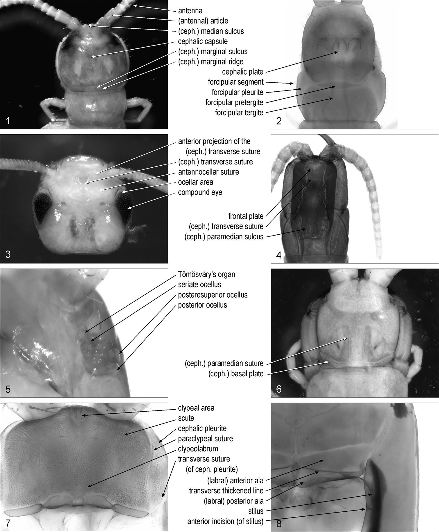
cephalic capsule: integument of the head to the exclusion of its appendages. Fig. 1. Syn.: head capsule
cephalic plate: [Cra, Dev, Geo, Lit, Sco] dorsal side of the cephalic capsule. Fig. 2. Syn.: cephalic shield, head plate, head shield
(cephalic) median sulcus: [Lit, Scu] mid-longitudinal sulcus on the anterior part of the cephalic capsule. Fig. 1. Syn.: (cephalic) median furrow
(cephalic) transverse suture: transverse suture on the anterior part of the dorsal side of the cephalic capsule. Figs 3–4. Syn.: cephalic suture, frontal line, frontal sulcus, frontal suture
anterior projection/projections of the (cephalic) transverse suture: [Scu] one of the paramedian sutures projecting anteriorly from the cephalic transverse suture. Fig. 3
antennocellar suture/sutures: [Cra, Lit, Scu] one of the paired sutures on the antero-lateral parts of the cephalic capsule. Fig. 3. Crabill 1961a: 131
antennal branch/branches of antennocellar suture: [Cra, Lit, Scu] part of the antennocellar suture, anterior to the cephalic transverse suture. Syn.: anterior portion/portions of antennocellar suture
ocellar branch/branches of antennocellar suture: [Cra, Lit, Scu] part of the antennocellar suture, posterior to the cephalic transverse suture. Syn.: posterior portion/portions of antennocellar suture, posterior limb/limbs of (cephalic) transverse suture
frontal plate: anterior part of the dorsal side of the cephalic capsule, delimited posteriorly by the cephalic transverse suture. Fig. 4. Syn.: frons
ocellar area/areas: [Lit, Scu] one of the paired antero-lateral parts of the cephalic capsule, bearing compound eyes or ocelli when present, and delimited mesally by the antennocellar suture. Fig. 3. Syn.: eye area/areas, ocellary area/areas, ocellary field/fields, ocular area/areas
compound eye/eyes: [Scu] faceted vision organ, composed of similar units known as ommatidia. Fig. 3
ocellus/ocelli: [Cra, Lit, Sco] simple vision organ, appearing as a single convex lens. Fig. 5
posterior ocellus/ocelli: [Lit] the most posterior ocellus on each side of the head. Fig. 5. Crabill 1961a: 132. Syn.: major ocellus/ocelli, principal ocellus/ocelli, terminal ocellus/ocelli
seriate ocellus/ocelli: [Lit] one of the ocelli other than the posterior ocellus. Fig. 5. Crabill 1961a: 132. Syn.: minor ocellus/ocelli
ocellar series/series: [Lit] one of the sub-horizontal rows in which the seriate ocelli can be arranged. Syn.: ocellar row/rows
posterosuperior ocellus/ocelli: [Lit] the most posterior ocellus of the most dorsal row of seriate ocelli. Fig. 5
Tömösváry’s organ/organs: [Cra, Lit, Scu] hygroreceptor sensory organ at the side of the head. Fig. 5. Syn.: organ/organs of Tömösváry, postantennal organ/organs, Tömösváry organ/organs
(cephalic) paramedian suture/sutures: [Sco] one of the paired paramedian sutures on the cephalic plate. Fig. 6
(cephalic) paramedian sulcus/sulci: [Geo, Lit] one of the paired paramedian sulci on the posterior part of the cephalic plate. Fig. 4. Syn.: paired posterior depression/depressions.
(cephalic) marginal ridge: [Lit, Sco] narrow ridge along the lateral and posterior margins of the dorsal side of the cephalic capsule. Fig. 1. Crabill 1961a: 131; Lewis et al. 2005: 7. Syn.: limbus, marginal bulge, marginal rim
(cephalic) marginal sulcus/sulci: [Lit, Sco] sulcus between the marginal ridge and the remaining part of the dorsal side of the cephalic capsule. Fig. 1
lateral marginal interruption/interruptions (of cephalic plate): [Lit] notch on the lateral margins of the cephalic plate. Eason 1964: 179. Syn.: disjuncture/disjunctures of limbus, lateral termination/terminations of marginal ridge
(cephalic) basal plate/plates: [Sco] one of the paired sclerites at the posterior corners of the cephalic plate. Fig. 6
cephalic pleurite/pleurites: [Cra, Dev, Geo, Lit, Sco] one of the pleurites lateral to the clypeolabrum. Fig. 7. Syn.: bucca/buccae, cephalic pleura/pleurae, cephalic pleuron/pleura
transverse suture/sutures (of cephalic pleurite): [Geo] transverse suture on the cephalic pleurite. Fig. 7. Crabill 1960b: 189. Syn.: buccal suture/sutures, transbuccal suture/sutures
stilus/stili: [Geo] sclerotised ridge on the mesal margin of the cephalic pleurite. Fig. 8. Crabill 1959a: 192; 1964a: 168; 1970: 235. Syn.: buccal margin/margins
anterior incision/incisions (of stilus): [Geo] notch on the mesal side of the stilus. Fig. 8. Crabill 1959a: 192
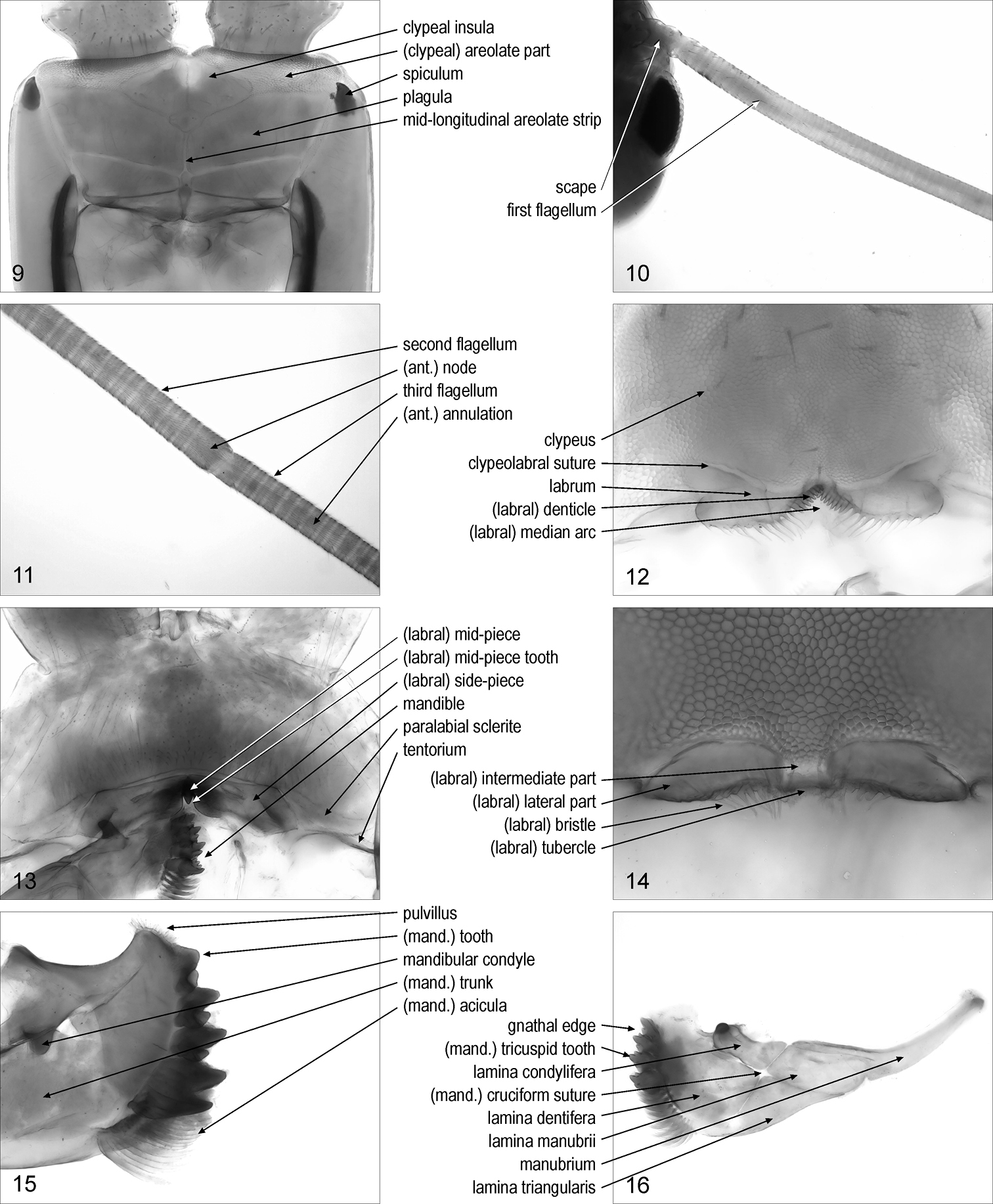
spiculum/spicula: [Geo] sclerotised, pointed projection on the anterior part of the cephalic pleurite. Fig. 9. Crabill 1959a: 192; 1964a: 168; 1970: 236
maxillary complex: whole of first and second maxillae
1 anterior part of body, dorsal, Lamyctes emarginatus 2 anterior part of body, dorsal, Geophilus carpophagus 3 head, dorsal, Scutigera coleoptrata 4 anterior part of body, dorsal, Mecistocephalus guildingii 5 left part of cephalic capsule, ventro-lateral, Lithobius dentatus 6 anterior part of body, dorsal, Cormocephalus gervaisianus 7 anterior part of cephalic capsule, without maxillary complex and mandibles, ventral, Ribautia centralis 8 left part of cephalic capsule, without maxillary complex and mandibles, ventral, Mecistocephalus togensis. Abbreviations: ceph., cephalic.
1 anterior part of body, dorsal, Lamyctes emarginatus 2 anterior part of body, dorsal, Geophilus carpophagus 3 head, dorsal, Scutigera coleoptrata 4 anterior part of body, dorsal, Mecistocephalus guildingii 5 left part of cephalic capsule, ventro-lateral, Lithobius dentatus 6 anterior part of body, dorsal, Cormocephalus gervaisianus 7 anterior part of cephalic capsule, without maxillary complex and mandibles, ventral, Ribautia centralis 8 left part of cephalic capsule, without maxillary complex and mandibles, ventral, Mecistocephalus togensis. Abbreviations: ceph., cephalic.
antenna/antennae: one of the paired most anterior appendages on the head. Fig. 1
(antennal) article/articles: one of the rigid sectors along the antenna. Fig. 1. Syn.: (antennal) annulus/annuli, (antennal) joint/joints, (antennal) segment/segments, antennomere/antennomeres
scape/scapes: [Scu] set of the two most basal antennal articles. Fig. 10
(antennal) annulation/annulations: [Scu] short antennal article. Fig. 11. Syn.: (antennal) article/articles
flagellum/flagella: [Scu] one of the sections along the antenna composed of annulations. Figs 10–11. Syn.: duploflagellum/duploflagella
(antennal) node/nodes: [Scu] elongate antennal article between two flagella along the antenna. Fig. 11
first flagellum/flagella: [Scu] the most basal flagellum along the antenna. Fig. 10. Syn.: flagellum/flagella primum/prima, first division/divisions of antenna/antennae
second flagellum/flagella: [Scu] the second flagellum along the antenna. Fig. 11. Syn.: flagellum/flagella secundum/secunda, second division/divisions of antenna/antennae
third flagellum/flagella: [Scu] the third flagellum along the antenna. Fig. 11. Syn.: flagellum/flagella tertium/tertia, third division/divisions of antenna/antennae
shaft organ/organs: [Scu] sensory organ on the first antennal article
9 anterior part of cephalic capsule, without maxillary complex and mandibles, ventral, Mecistocephalus togensis 10 basal part of right antenna, dorsal, Scutigera coleoptrata 11 intermediate part of the right antenna, dorsal, Scutigera coleoptrata 12 anterior part of cephalic capsule, without maxillary complex and mandibles, ventral, Pectiniunguis ducalis 13 anterior part of the cephalic capsule, without maxillary complex and left mandible, ventral, Scolopendra oraniensis 14 labrum, ventral, Ribautia centralis 15 distal part of right mandible, antero-dorsal, Lithobius dentatus 16 left mandible, antero-dorsal, Scolopendra oraniensis. Abbreviations: ant., antennal; mand., mandibular.
9 anterior part of cephalic capsule, without maxillary complex and mandibles, ventral, Mecistocephalus togensis 10 basal part of right antenna, dorsal, Scutigera coleoptrata 11 intermediate part of the right antenna, dorsal, Scutigera coleoptrata 12 anterior part of cephalic capsule, without maxillary complex and mandibles, ventral, Pectiniunguis ducalis 13 anterior part of the cephalic capsule, without maxillary complex and left mandible, ventral, Scolopendra oraniensis 14 labrum, ventral, Ribautia centralis 15 distal part of right mandible, antero-dorsal, Lithobius dentatus 16 left mandible, antero-dorsal, Scolopendra oraniensis. Abbreviations: ant., antennal; mand., mandibular.
clypeolabrum: antero-ventral part of the cephalic capsule, posterior to the antennae and between the cephalic pleurites. Fig. 7
clypeus: sclerite on the antero-ventral part of the cephalic capsule, to the exclusion of the labrum. Fig. 12
paraclypeal suture/sutures: one of the lateral margins of the clypeus. Fig. 7. Crabill 1959a: 192; 1959b: 173; 1960b: 189. Syn.: clypeal suture/sutures
scute/scutes: area on the cuticle, corresponding to the external face of a single epithelial cell. Fig. 7. Syn.: cuticular polygon/polygons
(clypeal) areolate part: [Geo] anterior part of the clypeus that is evidently areolate. Fig. 9. Syn.: areolate clypeus
plagula/plagulae: [Geo] one of the non-areolate areas on the posterior part of the clypeus. Fig. 9. Crabill 1959a: 192; 1959b: 173; 1964a: 168; 1970: 235. Syn.: (clypeal or prelabral) consolidated area/areas, (clypeal or prelabral) non-areolate field/fields, (clypeal or prelabral) non-areolate part/parts, posterior clypeus
mid-longitudinal areolate strip: [Geo] mid-longitudinal areolate band separating two paired plagulae. Fig. 9. Syn.: mid-longitudinal areolate stripe
clypeal insula/insulae: [Geo] non-areolate area inside the areolate part of the clypeus. Fig. 9
clypeal area/areas: [Geo] small, subcircular, median area on the areolate part of the clypeus, with distinctly finer or indistinct areolation. Fig. 7. Syn.: anteroclypeal area/areas, clypeal spot/spots, (clypeal or anterocentral) fenestra/fenestrae
clypeolabral suture: suture between clypeus and labrum. Fig. 12
labrum: posterior part of the clypeolabrum, sometimes delimited from the clypeus by a suture. Fig. 12. Syn.: upper lip
(labral) mid-piece: median sclerite of the labrum. Fig. 13. Syn.: (labral) median piece, (labral) middle piece
(labral) intermediate part: [Geo] median part of the labrum, when not a sclerite distinct from the lateral parts. Fig. 14. Syn.: median labromere, (labral) median piece, (labral) middle piece, (labral) middle portion, (labral) mid-piece
(labral) mid-piece tooth: sclerotised tooth on the labral mid-piece. Fig. 13
(labral) side-piece/side-pieces: one of the paired lateral sclerites of the labrum. Fig. 13. Syn.: (labral) lateral piece/pieces, (labral) lateral portion/portions
(labral) lateral part/parts: [Geo] one of the paired lateral parts of the labrum, when not sclerites distinct from the intermediate part. Fig. 14. Syn.: (labral) lateral portion/portions, (labral) side-piece/side-pieces
(labral) ala/alae: [Geo] one of the two sclerites composing the labral side-piece. Fig. 8
(labral) anterior ala/alae: [Geo] the anterior of the two sclerites composing the labral side-piece. Fig. 8
(labral) posterior ala/alae: [Geo] the posterior of the two sclerites composing the labral side-piece. Fig. 8
(labral) transverse thickened line/lines: [Geo] sclerotised ridge between the anterior and posterior ala of the labral side-piece. Fig. 8
(labral) median arc: [Geo] concave posterior margin of the labral intermediate part. Fig. 12. Syn.: labral arch
(labral) bristle/bristles: hair-like, sometimes branching, projection along the posterior margin of the labrum. Fig. 14. Syn.: (labral) branching bristle/bristles, (labral) filament/filaments, (labral) (branched) fimbria/fimbriae, (labral) hair/hairs
(labral) denticle/denticles: [Geo] subtriangular, flat projection along the posterior margin of the labrum. Fig. 12. Syn.: (labral) tooth/teeth
(labral) tubercle/tubercles: [Geo] subconical, stout projection along the posterior margin of the labrum. Fig. 14. Syn.: (labral) tooth/teeth
paralabial sclerite/sclerites: [Lit, Sco] one of the paired sclerites posterior to the clypeus and lateral to the labral side-pieces. Fig. 13. Syn.: coclypeus/coclypei
tentorium/tentoria: Y-shaped sclerite whose three limbs are attached to the labral lateral parts, the cephalic pleurite, and the mandibular condyle, respectively. Fig. 13. Syn.: (labral or mandibular) fulcrum/fulcra, (labral or labial) fultura/fulturae, tentorial complex/complexes
mandiblemandible/mandibles: one appendage of the first pair of the mouth-parts. Fig. 13
mandibular condyle/condyles: condyle of the mandible serving the articulation with the tentorium. Fig. 15
gnathal edge/edges: distal margin of the mandible. Fig. 16. Syn.: apical ridge/ridges, gnathal lobe/lobes, molar edge/edges
manubrium/manubria: slender projection of the mandible opposite to the gnathal edge with respect to the mandibular condyle. Fig. 16. Crabill 1960a: 15. Syn.: shank/shanks
(mandibular) trunk/trunks: main part of the mandible, to the exclusion of the manubrium and the gnathal edge. Fig. 15. Syn.: (mandibular) body/bodies, (mandibular) corpus/corpora, (mandibular) shaft/shafts
(mandibular) cruciform suture/sutures: [Sco] pair of crossed sutures on the mandibular trunk. Fig. 16. Syn.: cruciform fissure/fissures
lamina/laminae manubrii: [Sco] part of the mandible between manubrium and cruciform suture. Fig. 16. Crabill 1960a: 15
lamina/laminae triangularis/triangulares: [Sco] part of the mandible between the lamina manubrii and the lamina dentifera, opposite to the lamina condylifera with respect to the cruciform suture. Fig. 16. Crabill 1960a: 15
lamina/laminae dentifera/dentiferae: [Sco] part of the mandible between apical ridge and cruciform suture. Fig. 16. Crabill 1960a: 15
lamina/laminae condylifera/condyliferae: [Sco] part of the mandible between the lamina manubrii and the lamina dentifera, including the mandibular condyle and opposite to the lamina triangularis with respect to the cruciform suture. Fig. 16. Crabill 1960a: 15
molar plate/plates: [Scu] sclerotised, flat area on the gnathal edge
pulvillus/pulvilli: array of dense short scales on the dorsal end of the mandibular gnathal edge. Fig. 15. Syn.: furry pad/pads, Haarpolster
(mandibular) acicula/aciculae: one of the slender long projections on the ventral end of the mandibular gnathal edge. Fig. 15. Edgecombe 2001: 203. Syn.: sickle bristle/bristles, sickle-shaped bristle/bristles
pinnule/pinnules (of acicula): one of the branches of a mandibular acicula
(mandibular) branching bristle/bristles: [Lit] hair-like, branching projection fringing the mandibular teeth and aciculae
(mandibular) accessory denticle/denticles: [Lit, Sco] one of the denticles arranged in rows on the mandibular teeth
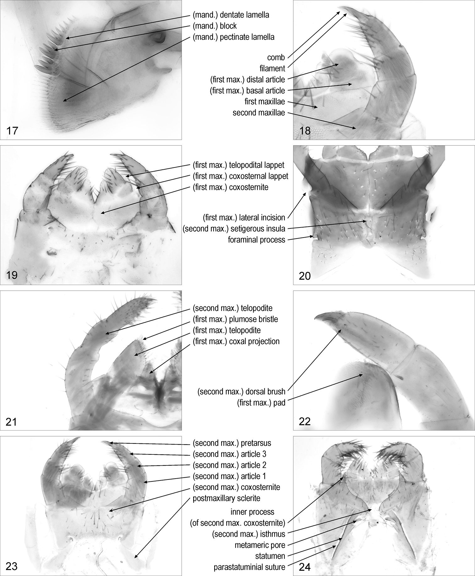
(mandibular) lamella/lamellae: one of the flat projections on the gnathal edge of the mandible. Fig. 17. Syn.: (mandibular) lamina/laminae
(mandibular) dentate lamella/lamellae: mandibular lamella bearing teeth. Fig. 17. Syn.: dentate lamina/laminae, dentate plate/plates, lamella/lamellae dentata/dentatae
(mandibular) tooth/teeth: sclerotised, large, subconical, projection on a mandibular dentate lamella. Fig. 15
(mandibular) tricuspid tooth/teeth: tooth with three tips on a dentate lamella. Fig. 16
(mandibular) block/blocks: one of the sclerotised distinct parts of a dentate lamella, each bearing one or more teeth. Fig. 17
(mandibular) pectinate lamella/lamellae: [Geo, Sco] mandibular lamella bearing poorly sclerotised, subcylindrical, slender projections. Fig. 17. Syn.: pectinate lamina/laminae
(mandibular) basal tooth/teeth: [Geo] subconical projection at the base of the first mandibular lamella
17 distal part of right mandible, posterior, Pectiniunguis ducalis 18 left part of maxillary complex, ventral, Pectiniunguis ducalis 19 maxillary complex, ventral, Geophilus carpophagus 20 maxillary complex, ventral, Mecistocephalus togensis 21 right part of maxillary complex, ventral, Lithobius dentatus 22 anterior right part of maxillary complex, dorsal, Scolopendra oraniensis 23 maxillary complex, ventral, Pectiniunguis ducalis 24 maxillary complex, ventral, Ribautia centralis. Abbreviations: mand., mandibular; max., maxillary.
17 distal part of right mandible, posterior, Pectiniunguis ducalis 18 left part of maxillary complex, ventral, Pectiniunguis ducalis 19 maxillary complex, ventral, Geophilus carpophagus 20 maxillary complex, ventral, Mecistocephalus togensis 21 right part of maxillary complex, ventral, Lithobius dentatus 22 anterior right part of maxillary complex, dorsal, Scolopendra oraniensis 23 maxillary complex, ventral, Pectiniunguis ducalis 24 maxillary complex, ventral, Ribautia centralis. Abbreviations: mand., mandibular; max., maxillary.
first maxillae {plural only}: pair of appendages and associated basal sclerites between the mandibles and the second maxillae. Fig. 18. Syn.: first maxilla {singular}, maxillae I {plural}
(first maxillary) sternite: most basal part of the coxosternite, associated with the first maxillae
(first maxillary) coxa/coxae: part of the coxosternite corresponding to a coxa, of the first maxillae
(first maxillary) coxosternite: entire sclerite corresponding to sternite and coxae of the first maxillae. Fig. 19. Syn.: coxae {plural}, coxites {plural}, coxosterna {singular}, coxosternum, sternum, syncoxite, syncoxosternum
(first maxillary) lateral incision: [Geo] notch on the lateral margin of the first maxillary coxosternite. Fig. 20. Crabil 1959a: 192
(first maxillary) coxal projection/projections: one of the paired projections on the anterior margin of the first maxillary coxosternite, mesal to the telopodites. Fig. 21. Syn.: coxal process/processes, inner branch/branches, inner lobe/lobes, medial lobe/lobes, medial projection/projections
(first maxillary) telopodite/telopodites: one of the paired projections, usually articulated at the base, on the anterior margin of the first maxillary coxosternite, lateral to the coxal projections. Fig. 21. Syn.: outer branch/branches, outer lobe/lobes, palp/palps, palpus/palpi
(first maxillary) basal article/articles: the most basal article of the first maxillary telopodite. Fig. 18. Syn.: femoroid/femoroids
(first maxillary) distal article/articles: the most distal article of the first maxillary telopodite. Fig. 18. Syn.: tibio-tarsus/tibio-tarsi
(first maxillary) plumose bristle/bristles: [Lit] one of the feather-like projections on the distal article of the first maxillary telopodite. Fig. 21
(first maxillary) pad/pads: [Sco] array of short, dense projections on the distal article of the first maxillary telopodite. Fig. 22
(first maxillary) lappet/lappets: [Geo] projection on the lateral margin of the first maxillary coxosternite or telopodite. Fig. 19. Syn.: external sensory lappet/lappets, (lateral or maxillary) palp/palps, palpal process/processes, (lateral or maxillary) palpus/palpi
(first maxillary) coxosternal lappet/lappets: [Geo] lappet on the first maxillary coxosternite. Fig. 19. Syn.: coxal palpus/palpi, syncoxal lobe/lobes, syncoxital lappet/lappets
(first maxillary) telopodital lappet/lappets: [Geo] lappet on the basal article of the first maxillary telopodite. Fig. 19. Syn.: femural palpus/palpi
second maxillaesecond maxillae {plural only}: pair of appendages and associated basal sclerite/s, posterior to the first maxillae. Fig. 18. Syn.: labium, maxillae II {plural}, second maxilla {singular}
(second maxillary) coxosternite: entire sclerite corresponding to sternite and coxae of the second maxillae. Fig. 23. Syn.: (second maxillary) coxosterna {singular}, (second maxillary) coxosternum
(second maxillary) isthmus: median part of the second maxillary coxosternite. Fig. 24
(second maxillary) setigerous insula/insulae: [Geo] one of the nonareolate areas, bearing setae, inside the areolate part of the second maxillary coxosternite. Fig. 20
metameric pore/pores: one of the paired pores of the maxillary glands on the second maxillary coxosternite. Fig. 24. Syn.: salivary pore/pores
foraminal process/processes: [Geo] marginal projection of the second maxillary coxosternite surrounding the metameric pore. Fig. 20
statumen/statuminia: [Geo] sclerotised elongated ridge mesal to the metameric pore. Fig. 24. Crabill 1955: 222; 1960b: 194. Syn.: (second maxillary) pleurocoxal line/lines, (second maxillary) pleurosternal suture/sutures
parastatuminial suture/sutures: [Geo] suture along the statumen. Fig. 24. Crabill 1964b: 39
circumforaminal ring/rings: [Geo] sclerotised ring partially surrounding the metameric pore
inner process/processes (of second maxillary coxosternite): [Geo] one of the paired projections on the anterior margin of the second maxillary coxosternite, mesal to the telopodites. Fig. 24. Syn.: (second maxillary) mesodistal process/processes
postmaxillary sclerite/sclerites: [Cra, Geo, Lit] one of the paired sclerites adjacent to the posterior corners of the second maxillary coxosternite. Fig. 23. Crabill 1960b: 189.
(second maxillary) telopodite/telopodites: part of the appendage of the second maxillae, distal to the most basal articulation. Fig. 21. Syn.: palp/palps, palpus/palpi, telopod/telopods
(second maxillary) trochanter/trochanters: [Scu] first article of the second maxillary telopodite. Fig. 25
(second maxillary) prefemur/prefemora: [Scu] second article of the second maxillary telopodite. Fig. 25
(second maxillary) femur/femora: [Scu] third article of the second maxillary telopodite. Fig. 25
(second maxillary) tibia/tibiae: [Scu] fourth article of the second maxillary telopodite. Fig. 25
(second maxillary) tarsus/tarsi: [Scu] fifth article of the second maxillary telopodite. Fig. 25
(second maxillary) article/articles 1: [Cra, Geo, Lit, Sco] first article of the second maxillary telopodite. Fig. 23. Lewis et al. 2005: 2, 3. Syn.: basal article/articles, femoroid/femoroids, first article/articles, telomere/telomeres 1
(second maxillary) article/articles 2: [Cra, Geo, Lit, Sco] second article of the second maxillary telopodite. Fig. 23. Lewis et al. 2005: 2, 3. Syn.: second article/articles, second joint/joints, telomere/telomeres 2, tibia/tibiae
(second maxillary) article/articles 3: [Cra, Geo, Lit, Sco] third article of the second maxillary telopodite. Fig. 23. Lewis et al. 2005: 2, 3. Syn.: apical article/articles, tarsus/tarsi, telomere/telomeres 3, terminal joint/joints, third article/articles, ultimate article/articles
(second maxillary) plumose seta/setae: [Lit] one of the setae with apical branches, on article 3 of the second maxillary telopodite. Fig. 26
(second maxillary) dorsal brush/brushes: [Cra, Sco] longitudinal row of hairs on article 3 of the second maxillary telopodite. Fig. 22. Lewis et al. 2005: 2, 3. Syn.: palisade/palisades of capitate hairs
(second maxillary) pretarsus/pretarsi: [Cra, Geo, Lit, Sco] terminal element articulated to the most distal article of the second maxillary telopodite. Fig. 23. Syn.: praetarsus/praetarsi
(second maxillary) claw/claws: [Cra, Geo, Lit, Sco] second maxillary pretarsus in shape of a claw. Fig. 26. Syn.: apical claw/claws, pretarsal claw/claws, terminal claw/claws
digit/digits (of second maxillary claw): [Cra, Lit] one of the short projections on the second maxillary claw. Fig. 26
comb/combs (of second maxillary claw): [Geo, Sco] row of projections along the margin of the second maxillary claw. Fig. 18. Syn.: comb/combs of teeth
filament/filaments (of second maxillary claw): [Geo, Sco] one of the slender projections of the comb of the second maxillary claw. Fig. 18
25 left part of second maxillae, ventral, Scutigera coleoptrata 26 distal part of second maxillary right telopodite, dorsal, Lithobius dentatus 27 forcipular segment, dorsal, Mecistocephalus togensis 28 anterior part of body, ventral, Lamyctes emarginatus 29 forcipular segment, ventral, Scutigera coleoptrata 30 forcipular segment, ventral, Clinopodes trebevicensis 31 anterior part of forcipular segment, ventral, Lithobius dentatus 32 right part of forcipular segment, ventral, Scolopendra oraniensis. Abbreviations: coxost., coxosternal; forc., forcipular; max., maxillary; troch., trochanteroprefemur.
25 left part of second maxillae, ventral, Scutigera coleoptrata 26 distal part of second maxillary right telopodite, dorsal, Lithobius dentatus 27 forcipular segment, dorsal, Mecistocephalus togensis 28 anterior part of body, ventral, Lamyctes emarginatus 29 forcipular segment, ventral, Scutigera coleoptrata 30 forcipular segment, ventral, Clinopodes trebevicensis 31 anterior part of forcipular segment, ventral, Lithobius dentatus 32 right part of forcipular segment, ventral, Scolopendra oraniensis. Abbreviations: coxost., coxosternal; forc., forcipular; max., maxillary; troch., trochanteroprefemur.
forcipular segment: segment bearing the forcipules. Fig. 2. Syn.: maxillipede segment, prehensorial segment
forcipular pretergite: [Geo, Sco] short sclerite anterior to the forcipular tergite. Fig. 2. Syn.: lamina basalis, prebasal plate
forcipular tergite: main tergite of the forcipular segment. Fig. 2. Syn.: basal plate
forcipular pleurite/pleurites: lateral sclerite of the forcipular segment. Fig. 2. Syn.: (forcipular or maxillipede) pleuron/pleura, (forcipular or maxillipede) pleura/pleurae
(forcipular) scapula/scapulae: [Geo] dorsal ridge of the forcipular pleurite. Fig. 27
(forcipular) scapular point/points: [Geo] projecting anterior tip of the forcipular scapula. Fig. 27. Crabill 1970: 237. Syn.: scapular projection/projections
(forcipular) collar: [Lit] ventral transversal bridge connecting the forcipular pleurites. Fig. 28. Syn.: (maxillipede) (pleural) collar
(forcipular) coxa/coxae: [Scu] one of the paired sclerites basal to the forcipules, bearing spine-bristles on the anterior margin. Fig. 29. Syn.: (forcipular) coxite/coxites
(forcipular) coxosternite: [Cra, Dev, Geo, Lit, Sco] entire sclerite corresponding to sternite and coxae of the forcipular segment. Fig. 28. Syn.: (maxillipede) coxosternite, (forcipular or maxillipede) coxosternum, (prehensorial) pre-sternum, (prehensorial) prosternum
(forcipular) coxopleural suture/sutures: suture between the forcipular pleurite and the forcipular coxae or coxosternite. Fig. 30. Syn.: pleuroprosternal suture/sutures
coxosternal condyle/condyles: condyle of the forcipular coxa or coxosternite serving the articulation with the trochanteroprefemur. Fig. 30. Syn.: (coxofemoral or prehensorial) condyle/condyles
(coxosternal) cerrus/cerri: [Geo] one of the paired groups of setae on the dorsal side of the forcipular coxosternite. Fig. 27. Crabill 1970: 236
(coxosternal) condylar process/processes: [Geo] one of the paired projections of the forcipular coxosternite, close to the dorsal coxosternal condyles
shoulder/shoulders (of forcipular coxosternite): [Lit] one of the paired obtuse projections on the anterior margin of the forcipular coxosternite. Fig. 31. Syn.: coxal endite/endites, lateral prosternal prominence/prominences
(coxosternal) median diastema: [Cra, Dev, Geo, Lit, Sco] median concavity on the anterior margin of the forcipular coxosternite. Fig. 31. Syn.: median interval, median notch, median sinus
(coxosternal) tooth/teeth: [Cra, Dev, Lit, Sco] sclerotised, short, subconical projection on the anterior margin of the forcipular coxosternite. Fig. 32. Syn.: (coxosternal or prosternal) (anterior or anterocentral) denticle/denticles, (prosternal or forcipular) tooth/teeth, (marginal) tubercle/tubercles
(coxosternal) tooth-plate/tooth-plates: [Cra, Sco] one of the paired sclerotised, flat, teeth-bearing projections on the anterior margin of the forcipular coxosternite. Fig. 32. Lewis et al. 2005: 2, 3. Syn.: dental plate/plates, (coxosternal or prosternal) plate/plates, (coxosternal or prosternal) toothed anterior process/processes
(coxosternal) denticle/denticles: [Geo] one of the paired small, subconical projections on the anterior margin of the forcipular coxosternite. Fig. 30. Syn.: (coxosternal) tooth/teeth
porodont/porodonts: [Lit] one of the paired large setae usually placed lateral to the forcipular coxosternal teeth. Fig. 31. Crabill 1953: 119. Syn.: accessory spine/spines, ectal spine/spines, ectodont/ectodonts, lateral (prosternal) spine/spines, parodontal spine/spines, pseudoporodont/pseudoporodonts
porodont node/nodes: [Lit] basal structure from which the porodont arises
(coxosternal) median cleft: [Lit] mid-longitudinal suture on the ventral side of the forcipular coxosternite. Fig. 28. Eason 1964: 165.
chitin-line/chitin-lines: [Geo] one of the paired paramedian sclerotised narrow stripes on the ventral side of the forcipular coxosternite. Fig. 30. Crabill 1960a: 15.Syn.: chitinous line/lines, pleurogram/pleurograms, (prosternal) (subcondylic) sclerotic line/lines
(coxosternal) median embayment: [Lit, Sco] median notch at the posterior margin of the forcipular coxosternite. Fig. 28. Edgecombe and Koch 2008: 895
forcipule/forcipules: telopodite of the forcipular segment. Fig. 28. Lewis 1981: 11. Syn.: forcipula/forcipulae, forcipular telopodite/telopodites, maxilliped/maxillipeds, maxillipede/maxillipedes, poison claw/claws, prehensor/prehensors, prehensorial foot/feet, prehensorial telopodite/telopodites, toxicognath/toxicognaths
(forcipular) trochanteroprefemur/trochanteroprefemora: first article of the forcipule. Figs 29, 32. Syn.: article/articles 1, basal article/articles, femoroid/femoroids, femuroid/femuroids, first article/articles
(forcipular) trochanteral suture/sutures: trace of suture on the forcipular trochanteroprefemur. Fig. 29
(forcipular) intermediate article/articles: one of the second and third articles of the forcipule. Syn.: intercalary article/articles, intermediate joint/joints
(forcipular) femur/femora: second article of the forcipule. Figs 29, 32. Syn.: article/articles 2, femoroid/femoroids, femuroid/femuroids, second article/articles, second joint/joints, tibia/tibiae
(forcipular) tibia/tibiae: third article of the forcipule. Figs 29, 32. Syn.: article/articles 3, tarsus/tarsi, third article/articles, third joint/joints, tibioid/tibioids
(forcipular) tarsus/tarsi: [Scu] fourth article of the forcipule. Fig. 29(forcipular)
ungulum/ungula: [Scu] terminal articulated element of the forcipule. Fig. 29. Syn.: pretarsus/pretarsi, ungula/ungulae
(forcipular) tarsungulum/tarsungula: [Cra, Dev, Geo, Lit, Sco] ultimate article of the forcipule. Fig. 32. Lewis et al. 2005: 2. Syn.: article/articles 4, claw/claws, metatarsus/metatarsi, poison claw/claws, pretarsus/pretarsi, tarsungula/tarsungulae
process/processes of (forcipular) tronchanteroprefemur: [Cra, Sco] large projection on the mesal side of the forcipular trochanteroprefemur. Fig. 32. Lewis et al. 2005: 2, 3. Syn.: (tronchanteroprefemoral) (inner spinous) process/processes, process/processes of femoroid, (tronchanteroprefemoral) (inner or median) tooth/teeth
(forcipular) denticle/denticles: [Geo] small subconical projection on the mesal side of the forcipule. Figs 27, 30. Syn.: node/nodes, nodule/nodules, tooth/teeth
proximal denticle/denticles (of trochanteroprefemur): [Geo] the most basal of two denticles along the mesal side of the forcipular trochanteroprefemur. Fig. 27. Syn.: basal denticle/denticles (of trochanteroprefemur)
distal denticle/denticles (of trochanteroprefemur): [Geo] the most distal of two denticles along the mesal side of the forcipular trochanteroprefemur. Fig. 27
basal denticle/denticles (of tarsungulum): [Geo] denticle at the base of the forcipular tarsungulum. Fig. 30. Syn.: basal tooth/teeth, (basal) node/nodes
(forcipular) spine comb/combs: [Scu] row of spines on the forcipular tarsus
leg-bearing segmentleg-bearing segment/segments: segment of the trunk bearing paired walking appendages. Fig. 33. Syn.: pedal segment/segments, pedigerous (post-maxillipede) segment/segments, trunk-segment/trunk-segments
tergite/tergites (of leg-bearing segment): sclerite on the dorsal side of a leg-bearing segment. Fig. 34. Syn.: dorsal plate/plates, dorsal shield/shields, (dorsal) scutum/scuta, tergal plate/plates, tergum/terga {inappropriate use; see, e.g., Snodgrass 1935}
stomatotergite/stomatotergites: [Scu] tergite bearing a stoma. Fig. 35
stoma/stomata: [Scu] elongate opening of the respiratory organs on the posteromedian part of a stomatotergite. Fig. 35. Syn.: spiracle/spiracles, stigma/stigmata
stoma-saddle/stoma-saddles: [Scu] domed region of the stomatotergite surrounding the stoma. Fig. 35. Syn.: saddle/saddles
margination/marginations (of tergite): [Lit, Sco] marginal ridge on a tergite. Fig. 34. Lewis et al. 2005: 2. Syn.: border/borders, marginal ridge/ridges
posterior triangular projection/projections (of tergite): [Lit] angulated projection on each posterior corner of a tergite. Fig. 34. Syn.: angulation/angulations, posterior production/productions, posterior tergital projection/projections
pretergite/pretergites: [Geo, Sco] anterior sclerite of the two dorsal sclerites of a leg-bearing segment. Fig. 33. Syn.: intercalary tergite/tergites, intertergite/intertergites, prescutum/prescuta, pretergum/preterga, protergite/protergites
metatergite/metatergites: [Geo, Sco] posterior sclerite of the two dorsal sclerites of a leg-bearing segment. Fig. 33. Syn.: tergite/tergites
paramedian sulcus/sulci or suture/sutures (of tergite): [Geo, Sco] one of the paired paramedian longitudinal sutures or sulci on a tergite. Fig. 33. Lewis et al. 2005: 2, 3, 5. Syn.: paramedian groove/grooves, paramedian longitudinal sulcus/sulci or suture/sutures
(anterior) transverse sulcus/sulci or suture/sutures (of tergite): [Sco] transverse suture or sulcus on the first trunk tergite. Fig. 33. Lewis et al. 2005: 2, 3. Syn.: (procurved) cervical groove/grooves, (anterior) cervical sulcus/sulci or suture/sutures, semi-lunar sulcus/sulci, T1 ring suture/sutures, transversal suture/sutures, transverse collar sulcus/sulci, (procurved) transverse groove/grooves
cruciform suture/sutures (of tergite): [Sco] pair of crossed sutures on the first trunk tergite. Fig. 33
oblique suture/sutures (of tergite): [Sco] one of the paired oblique sutures on some anterior trunk tergites. Fig. 33. Lewis et al. 2005: 5. Syn.: arcuate suture/sutures, curved lateral sulcus/sulci
lateral longitudinal suture/sutures (of tergite): [Sco] one of the paired longitudinal sutures close to the lateral margins of a tergite. Fig. 33. Lewis et al. 2005: 5
lateral crescentic sulcus/sulci (of tergite): [Sco] one of the paired curved, sublongitudinal sulci on a tergite. Lewis et al. 2005: 5
eupleurium/eupleuria: the whole of pleurites on a side of a leg-bearing segment. Eason 1964: 277. Syn.: pleuron/pleura
pleural membrane/membranes: arthrodial membrane between pleurites
paratergite/paratergites: [Geo] pleurite contiguous or almost contiguous to a tergite. Fig. 36
intercalary paratergite/paratergites: [Geo] paratergite lateral to a pretergite. Fig. 36. Crabill 1960c: 93. Syn.: parapretergite/parapretergites, preparatergite/preparatergites, secondary paratergite/paratergites, suprascutellum/suprascutella
primary intercalary paratergite/paratergites: [Geo] the most mesal of two intercalary paratergites. Fig. 36. Syn.: primary suprascutellum/suprascutella
secondary intercalary paratergite/paratergites: [Geo] the most distal of two intercalary paratergites. Fig. 36. Syn.: secondary suprascutellum/suprascutella
principal paratergite/paratergites: [Geo] paratergite lateral to a metatergite. Fig. 36. Crabill 1960c: 93. Syn.: major paratergite/paratergites, primary paratergite/paratergites
scutellum/scutella: [Geo, Sco] pleurite in antero-ventral position with respect to the stigmatopleurite. Fig. 36. Syn.: intercalary pleurite/pleurites, prescutellum/prescutella
spiracle/spiracles: [Cra, Dev, Geo, Lit, Sco] one of the paired openings of the tracheae on the lateral sides of a leg-bearing segment. Fig. 36. Syn.: stigma/stigmata
stigmatopleurite/stigmatopleurites: [Cra, Dev, Geo, Lit, Sco] pleurite bearing a spiracle. Fig. 36. Syn.: spiracle-bearing pleurite/pleurites, spiraculiferous plate/plates, stigma-bearing pleurite/pleurites, stigmopleurite/stigmopleurites
catapleurite/catapleurites: pleurite between the scutellum and the coxa. Fig. 36. Syn.: catopleure/catopleures, katopleure/katopleures
eucoxa/eucoxae superior/superiores: [Lit] pleurite dorsal to the coxa
eucoxa/eucoxae inferior/inferiores: [Lit] pleurite ventral to the coxa
pleurocoxa/pleurocoxae: [Geo] pleurite between coxa and metacoxa
subcoxa/subcoxae: [Cra, Dev, Geo, Lit, Sco] pleurite anterior or posterior to the coxa. Syn.: subcoxal pleurite/pleurites
procoxa/procoxae: [Cra, Dev, Geo, Lit, Sco] subcoxa anterior to the coxa. Fig. 37. Syn.: anterior subcoxal plate/plates, precoxa/precoxae, procoxal pleurite/pleurites, prosubcoxa/prosubcoxae
metacoxa/metacoxae: [Cra, Dev, Geo, Lit, Sco] subcoxa posterior to the coxa. Fig. 37. Syn.: metacoxal pleurite/pleurites, metasubcoxa/metasubcoxae
sternite/sternites (of leg-bearing segment): sclerite on the ventral side of a leg-bearing segment. Fig. 38. Syn.: sternum/sterna {inappropriate use; see, e.g., Snodgrass, 1935}, ventral plate/plates, ventral shield/shields
presternite/presternites: [Cra, Dev, Geo, Sco] anterior region of the single sternite of a leg-bearing segment, or anterior sclerite of the two ventral sclerites of a leg-bearing segment. Fig. 37. Syn.: intercalary sternite/sternites, intersternite/intersternites, prosternite/prosternites
metasternite/metasternites: [Cra, Dev, Geo, Sco] posterior region of the single sternite of a leg-bearing segment, or posterior sclerite of the two ventral sclerites of a leg-bearing segment. Fig. 37. Syn.: sternite/sternites
transverse sulcus/sulci (of sternite): [Sco] transverse sulcus on a sternite. Fig. 39. Lewis et al. 2005: 5
median longitudinal sulcus/sulci (of sternite): [Sco] mid-longitudinal sulcus on a sternite. Fig. 39. Lewis et al. 2005: 5. Syn.: median sulcus/sulci, mid-longitudinal sulcus/sulci
cruciform suture/sutures (of sternite): [Sco] the pair of transverse and median longitudinal sulci on a sternite. Fig. 39. Syn.: cross furrow/furrows, cross sulcus/sulci, cruciform impression/impressions, cruciform sulcus/sulci
trigonal suture/sutures (of sternite): [Sco] pair of crossed sutures on the posterior part of the sternite. Lewis et al. 2005: 5
endosternite/endosternites: [Geo, Sco] posterior projection of a sternite, covered by the sternite of the following segment. Fig. 37. Syn.: metasternite/metasternites
carpophagus peg/pegs: [Geo] median projection on the posterior margin of a sternite, in the carpophagus-structure. Fig. 37. Crabill 1954: 174. Syn.: paxillus/paxilli
carpophagus pit/pits: [Geo] median socket on the anterior margin of a sternite, in the carpophagus-structure. Fig. 37. Crabill 1954: 174. Syn.: carpophagus fossa/fossae, sacculus/sacculi, sternal pit/pits
carpophagus-structure/carpophagus-structures: [Geo] whole of a carpophagus peg and the associated carpophagus pit. Fig. 37
ventral pore/pores: [Geo] glandular pore on the ventral side of a leg-bearing segment. Fig. 40. Syn.: sternal pore/pores, sternital pore/pores
(ventral) pore-field/pore-fields: [Geo] an area of clustered pores on the ventral side of a leg-bearing segment. Fig. 40. Syn.: pore area/areas, pore-group/pore-groups, poriferous area/areas, porigerous area/areas
sternobothrium/sternobothria: [Geo] median horseshoe-like pit on the metasternite. Fig. 40
transverse fossa/fossae (of sternite): [Geo] transverse, ellipical depression on some trunk sternites. Fig. 41. Eason 1964: 54
fungiform fovea/foveae: [Geo] median T-like pit on the metasternite
virguliform fossa/fossae: [Geo] comma-like pit at each of the anterior corners of a sternite. Fig. 42. Eason 1964: 284. Syn.: sternal pouch/pouches
lateral gutter/gutters (of sternite): [Geo] longitudinal groove along the lateral margin of a sternite. Fig. 42. Eason 1964: 48. Syn.: parasternital cleft/clefts, parasternital fossa/fossae, parasternital pit/pits
33 anterior part of trunk, dorsal, Cryptops anomalans 34 intermediate part of trunk, dorsal, Lithobius dentatus 35 intermediate part of trunk, dorsal, Scutigera coleoptrata 36 intermediate part of trunk, left, Himantarium gabrielis 37 intermediate part of trunk, ventral, Clinopodes trebevicensis 38 intermediate part of trunk, ventral, Cryptops parisi 39 intermediate part of trunk, ventral, Cryptops punicus 40 intermediate part of trunk, ventral, Bothriogaster signata.
33 anterior part of trunk, dorsal, Cryptops anomalans 34 intermediate part of trunk, dorsal, Lithobius dentatus 35 intermediate part of trunk, dorsal, Scutigera coleoptrata 36 intermediate part of trunk, left, Himantarium gabrielis 37 intermediate part of trunk, ventral, Clinopodes trebevicensis 38 intermediate part of trunk, ventral, Cryptops parisi 39 intermediate part of trunk, ventral, Cryptops punicus 40 intermediate part of trunk, ventral, Bothriogaster signata.
leg/legs: one of the paired appendages of the trunk to the exclusion of the forcipules and the gonopods. Fig. 41. Syn.: (ambulatory or locomotory or walking) leg/legs
cursiped/cursipeds: [Lit] a leg of the pairs 1–13. Crabill 1961a: 132
tenaciped/tenacipeds: [Lit] a leg of the pairs 14–15. Crabill 1961a: 132
(leg) article/articles: one of the articulated elements of a leg. Fig. 41. Syn.: podomere/podomeres, (leg) segment/segments
(leg) coxa/coxae: the most basal article of a leg. Fig. 38
(leg) trochanter/trochanters: small, basalmost article of a telopodite. Fig. 38
(leg) prefemur/prefemora: second article of a telopodite. Fig. 38. Syn.: praefemur/praefemora
(leg) femur/femora: third article of a telopodite. Fig. 43
(leg) tibia/tibiae: fourth article of a telopodite. Fig. 43. Syn.: patellotibia/patellotibiae
(leg) tarsus/tarsi: fifth article of a telopodite, when ultimate. Fig. 43
tarsal article/articles: one of the articles of a biarticulated region of the leg corresponding to the tarsus. Syn.: tarsalium/tarsalia, tarsomere/tarsomeres
tarsus/tarsi 1: the basal article of two tarsal articles. Fig. 44. Lewis et al. 2005: 2. Syn.: basitarsus/basitarsi, first division/divisions of tarsus/tarsi, first tarsal article/articles, first tarsal joint/joints, first tarsal segment/segments, first tarsus/tarsi, I tarsus/tarsi, protarsus/protarsi, proximotarsus/proximotarsi, tarsomere/tarsomeres 1, tarsus/tarsi, tarsus/tarsi I
tarsus/tarsi 2: the distal article of two tarsal articles. Fig. 44. Lewis et al. 2005: 2. Syn.: distitarsus/distitarsi, distotarsus/distotarsi, II tarsus/tarsi, metatarsus/metatarsi, pretarsus/pretarsi, second division/divisions of tarsus/tarsi, second tarsal article/articles, second tarsal joint/joints, second tarsal segment/segments, second tarsus/tarsi, tarsomere/tarsomeres 2, tarsus/tarsi II, telotarsus/telotarsi
(tarsal) annulation/annulations: [Lit, Sco, Scu] each part of a tarsal article, between two contiguous constrictions. Fig. 45. Syn.: annulus/annuli, pseudosegment/pseudosegments, secondary article/articles, tarsale/tarsalia, tarsomere/tarsomeres
carina/carinae: [Scu] longitudinal ridge on a leg article. Fig. 46. Edgecombe and Giribet 2006: 509
tarsal papilla/papillae: [Scu] relatively short, stout projection with a rounded tip, on the ventral side of tarsus 2. Fig. 45. Edgecombe and Giribet 2006: 509
resilient sole-hair/sole-hairs: [Scu] one of the paired hairs thickened at the base, on the ventral side of the leg, originating near the posteromedian margin of each tarsal papilla. Fig. 45. Würmli 1974: 95; Edgecombe and Giribet 2006: 512
(leg) spur/spurs: [Lit, Sco] spur on legs. Fig. 44. Syn.: calcar/calcars, (leg or pedal) spine/spines, spiniform seta/setae
distal spinose projection/projections (of tibia): [Lit] spinous process at the distal end of the tibia of a leg. Fig. 43. Syn.: tibial spur/spurs
pectinal seta/setae: [Lit] one of the decumbent setae arranged in rows along the tarsal articles of legs. Fig. 44. Crabill 1958: 262
(tarsal) pecten/pectines: [Lit] row of pectinal setae. Fig. 44. Crabill 1958: 262
pretarsus/pretarsi: apical element articulated at the tip of a leg. Fig. 43. Lewis et al. 2005: 2. Syn.: praetarsus/praetarsi, postarsus/postarsi, posttarsus/posttarsi
claw/claws: pretarsus in shape of a claw. Fig. 44. Lewis et al. 2005: 2. Syn.: apical claw/claws, end claw/claws, tarsal claw/claws
fundus/fundi (of claw): basal, swollen part of a claw. Fig. 47. Crabill 1961b: 501
unguis/ungues proper: distal, slender part of a claw. Fig. 47. Syn.: claw/claws proper, main claw/claws, principal claw/claws, unguis/ungues
accessory spine/spines: slender, pointed projection at the base of the claw. Fig. 47. Lewis et al. 2005: 2, 3. Syn.: accessory claw/claws, accessory seta/setae, accessory spur/spurs, basal bristle/bristles, basal spine/spines, basal spur/spurs, claw spine/spines, parunguis/parungues, sensory spine/spines, sensory spur/spurs
anterior accessory spine/spines: the anterior accessory spine of two of a claw. Fig. 47. Syn.: anterior accessory claw/claws, anterior accessory spur/spurs, anterior parunguis/parungues
posterior accessory spine/spines: the posterior accessory spine of two of a claw. Fig. 47. Syn.: posterior accessory claw/claws, posterior accessory spur/spurs, posterior parunguis/parungues
(pretarsal) posteroventral spine/spines: [Lit] the largest of a pair of spines emerging on the ventro-posterior side at the base of a claw, close to the posterior accessory spine. Edgecombe 2004: 31; Koch and Edgecombe 2008: 168
(pretarsal) subsidiary spine/spines: [Lit] the smallest of a pair of spines emerging on the ventro-posterior side at the base of a claw, close to the posterior accessory spine. Edgecombe 2004: 31
fibulunguis/fibulungues: [Geo] pretarsus with a large projection flanking the unguis proper. Fig. 48. Crabill 1969: 38
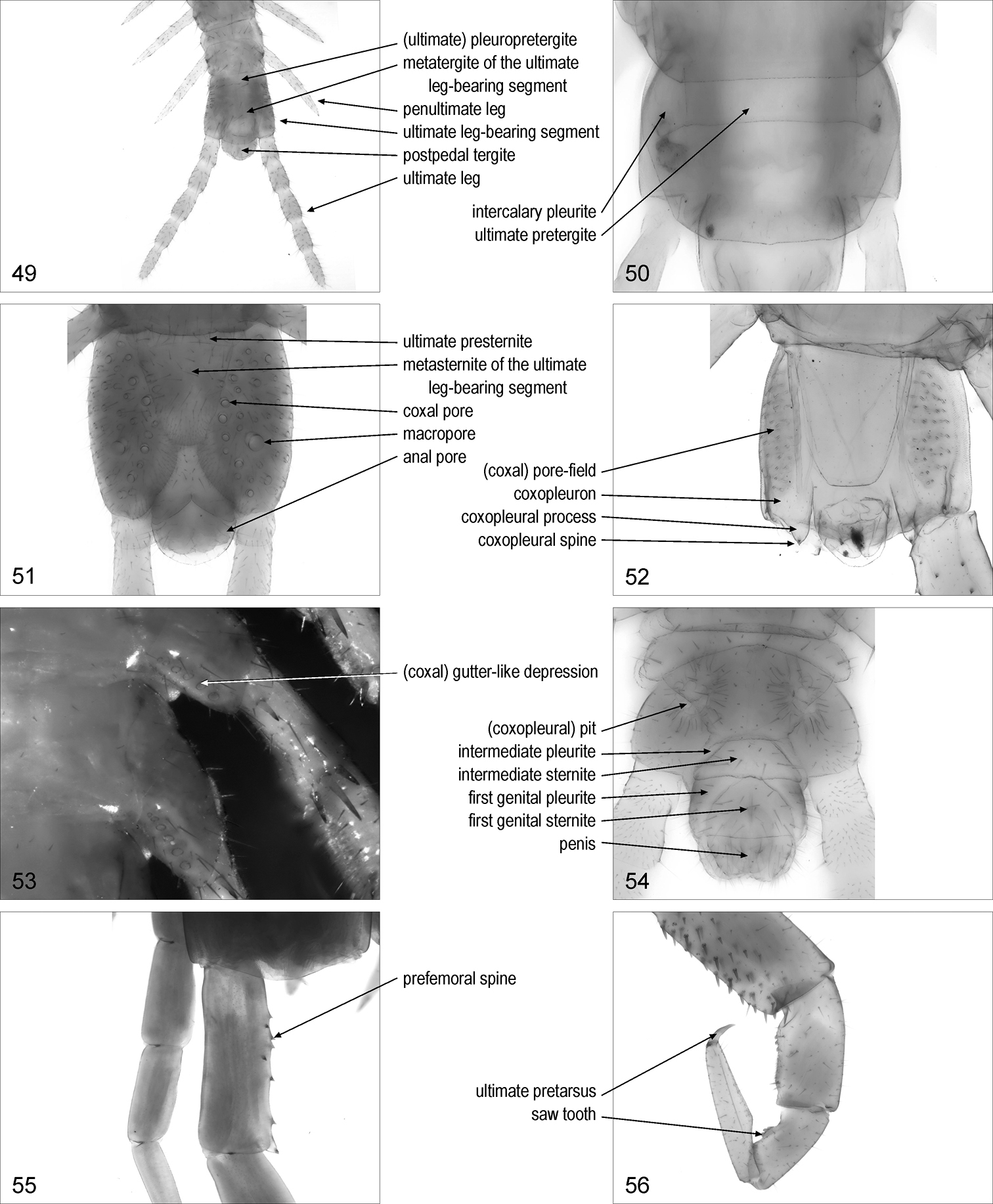
penultimate leg/legs: leg of the penultimate pair. Fig. 49. Syn.: penult leg/legs
41 intermediate part of trunk, ventral, Haplophilus souletinus 42 intermediate part of trunk, ventral, Stigmatogaster gracilis 43 right leg, anterior, Lamyctes emarginatus 44 distal part of right leg, antero-ventral, Lithobius dentatus 45 tarsus of left leg, anterior, Scutigera coleoptrata 46 intermediate part of left leg, anterior, Scutigera coleoptrata 47 distal part of left leg, ventral, Scolopendra oraniensis 48 distal part of left leg, anterior, Diphyonyx conjungens
41 intermediate part of trunk, ventral, Haplophilus souletinus 42 intermediate part of trunk, ventral, Stigmatogaster gracilis 43 right leg, anterior, Lamyctes emarginatus 44 distal part of right leg, antero-ventral, Lithobius dentatus 45 tarsus of left leg, anterior, Scutigera coleoptrata 46 intermediate part of left leg, anterior, Scutigera coleoptrata 47 distal part of left leg, ventral, Scolopendra oraniensis 48 distal part of left leg, anterior, Diphyonyx conjungens
ultimate leg-bearing segment: leg-bearing segment bearing the ultimate pair of legs. Fig. 49. Syn.: last leg-bearing segment, last pediferous segment, last trunk-segment, ultimate pedal segment, ultimate pedigerous segment
ultimate pretergite: [Geo] pretergite of the ultimate leg-bearing segment. Fig. 50
intercalary pleurite/pleurites: [Geo] pleurite contiguous to the ultimate pretergite. Fig. 50. Syn.: prepleurite/prepleurites
(ultimate) pleuropretergite: [Geo] entire sclerite corresponding to the ultimate pretergite and the two intercalary pleurites. Fig. 49
tergite (or metatergite) of the ultimate leg-bearing segment: main tergite of the ultimate leg-bearing segment. Fig. 49. Syn.: last dorsal plate, last tergite, ultimate tergite
ultimate presternite: [Geo] presternite of the ultimate leg-bearing segment. Fig. 51
sternite (or metasternite) of the ultimate leg-bearing segment: main sternite of the ultimate leg-bearing segment. Fig. 51. Syn.: last sternite, last sternum, last ventral plate, sternite (or metasternite) of the last trunk segment, sternite (or metasternite) of the last leg-bearing segment, ultimate (pedal) sternite
ultimate leg/legs: one of the legs of the ultimate pair. Fig. 49. Syn.: anal leg/legs, caudal leg/legs, end leg/legs, last leg/legs, posterior leg/legs, terminal leg/legs
coxopleuron/coxopleura: [Geo, Sco] basal element of the ultimate leg, corresponding to coxa and pleurites. Fig. 52. Lewis et al. 2005: 2. Syn.: (anal or last) coxa/coxae, coxopleura/coxopleurae, coxopleurite/coxopleurites, (last) pleura/pleurae, pleurocoxa/pleurocoxae
coxopleural process/processes: [Sco] posterior process of the coxopleuron. Fig. 52. Lewis et al. 2005: 2. Syn.: coxal process/processes, process/processes of last coxa/coxae
(coxopleural) spine/spines: [Sco] spine on the coxopleuron. Fig. 52. Lewis et al. 2005: 2, 5. Syn.: (coxopleural) spur/spurs
(coxopleural) side spine/spines: [Sco] spine on the posterior margin of the coxopleuron mesal to the coxopleural process. Lewis et al. 2005: 5
coxal pore/pores: [Cra, Dev, Geo, Lit, Sco] one of the pores of the coxal organs on posterior legs. Fig. 51. Syn.: coxopleural pore/pores, pleural pore/pores
macropore/macropores: [Geo] coxal pore that is distinctly larger than the other pores. Fig. 51
(coxal) gutter-like depression/depressions: [Lit] depressed area on the coxopleuron containing the openings of the coxal organs. Fig. 53
(coxopleural) pit/pits: [Geo] pit on the coxopleuron, containing the openings of the coxal organs. Fig. 54. Crabill 1961a: 133. Syn.: (coxal or coxopleural) crypt/crypts, gland pit/pits, subsurface gland-pit/gland-pits, subsurface pocket/pockets
(coxopleural) fossa/fossae: [Geo] longitudinal pouch close to the mesal margin of the coxopleuron, containing the openings of the coxal organs. Syn.: (coxopleural or porigerous) cavity/cavities, (coxopleural or porigerous) fossula/fossulae
(coxal) pore-field/pore-fields: [Cra, Dev, Geo, Lit, Sco] part of the surface of the coxa or coxopleuron of the ultimate legs containing the coxal pores. Fig. 52. Syn.: (coxal) cribriform area/areas, (coxal) porose area/areas
prefemoral spine/spines: [Sco] spine on the prefemur of ultimate and/or penultimate legs. Fig. 55. Lewis et al. 2005: 3, 5. Syn.: prefemoral dorsal spur/spurs, prefemoral tooth/teeth
prefemoral (spinous) process/processes: [Sco] process, usually bearing spines, on the prefemur of the ultimate legs. Lewis et al. 2005: 3, 5, 7. Syn.: distomedial prefemoral tubercle/tubercles(prefemoral)
corner spine/spines: [Sco] spine on the distal end, on the mesal side, of the prefemur of ultimate legs. Lewis et al. 2005: 3, 5. Syn.: (prefemoral) distomedial spine/spines
saw tooth/teeth: [Sco] one of the bluntly pointed projections arranged in rows on the tibia and tarsus 1 of ultimate legs. Fig. 56. Lewis et al. 2005: 7. Syn.: (tibial and tarsal) (serrate) comb/combs, mucro/mucrones, saw-like tooth/teeth
ultimate pretarsus/pretarsi: pretarsus of the ultimate leg. Fig. 56
49 terminal part of trunk, dorsal, Gnathoribautia bonensis 50 ultimate leg-bearing segment, dorsal, Bothriogaster signata 51 terminal part of trunk, ventral, female Dicellophilus carniolensis 52 terminal part of trunk, ventral, Cormocephalus gervaisianus 53 basal part of posterior left legs, ventral, Lithobius dentatus 54 terminal part of trunk, ventral, male Tuoba sydneyensis 55 basal part of ultimate left legs, dorsal, Scolopendra oraniensis 56 ultimate right leg, posterior, Cryptops anomalans.
49 terminal part of trunk, dorsal, Gnathoribautia bonensis 50 ultimate leg-bearing segment, dorsal, Bothriogaster signata 51 terminal part of trunk, ventral, female Dicellophilus carniolensis 52 terminal part of trunk, ventral, Cormocephalus gervaisianus 53 basal part of posterior left legs, ventral, Lithobius dentatus 54 terminal part of trunk, ventral, male Tuoba sydneyensis 55 basal part of ultimate left legs, dorsal, Scolopendra oraniensis 56 ultimate right leg, posterior, Cryptops anomalans.
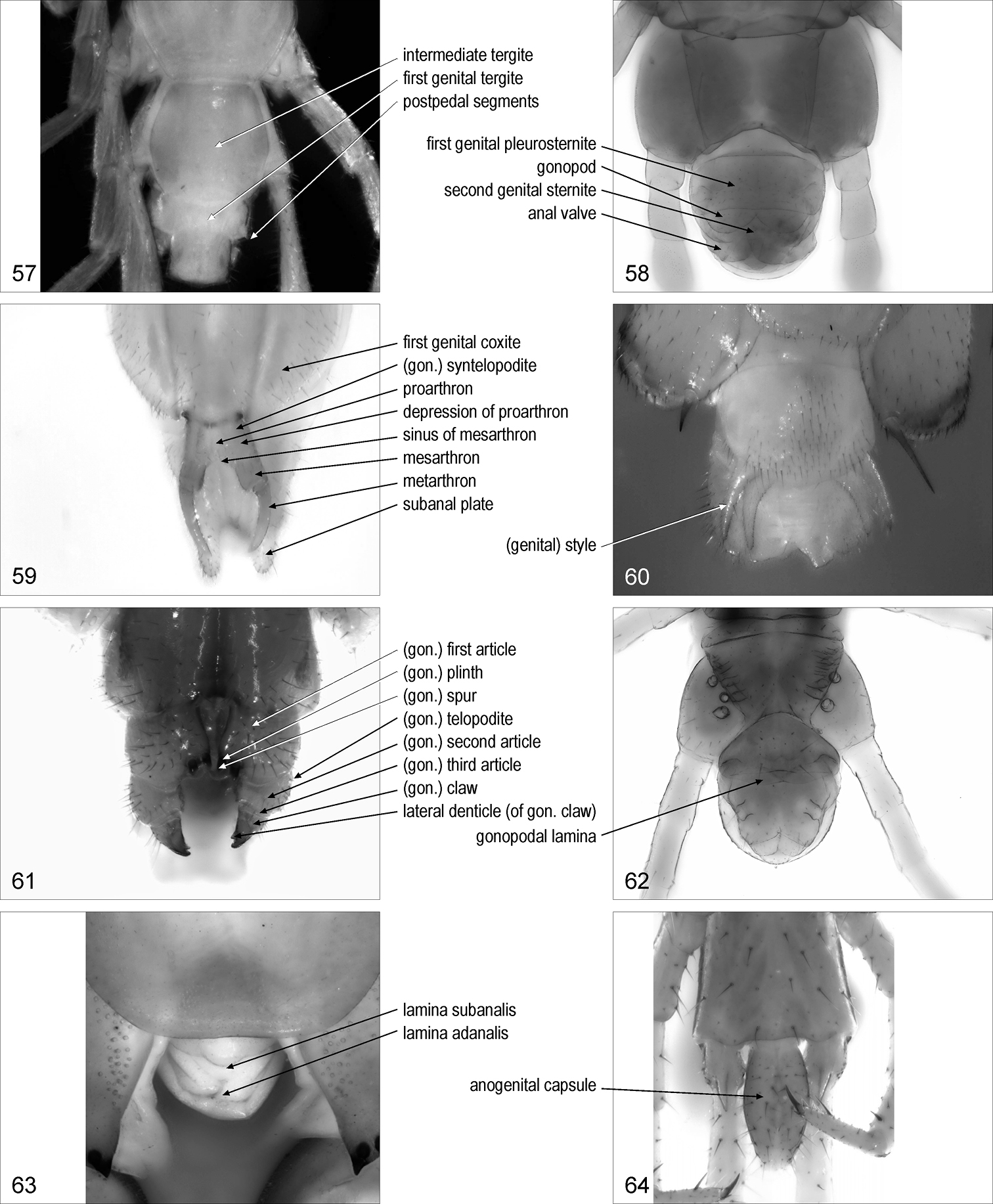
postpedal segments {plural only}: part of the trunk posterior to the ultimate leg-bearing segment. Fig. 57. Syn.: terminal segments
intermediate tergite: [Lit] tergite posterior to the tergite of the ultimate leg-bearing segment, corresponding to the intermediate sternite. Fig. 57. Eason 1964: 167
intermediate sternite: sternite between the sternite or metasternite of the ultimate leg-bearing segment and the first genital sternite. Fig. 54
intermediate pleurite/pleurites: [Geo] one of the two pleurites flanking the intermediate sternite. Fig. 54
first genital tergite: [Lit] tergite posterior to the intermediate tergite, corresponding to the first genital sternite. Fig. 57
first genital sternite: sternite between the intermediate sternite and the second genital sternite, usually associated with gonopods. Fig. 54. Syn.: pregenital sternite, sternite of first genital segment
first genital pleurite/pleurites: one of the two pleurites flanking the first genital sternite. Fig. 54. Syn.: pleurite of first genital segment
first genital pleurosternite: entire sclerite corresponding to the first genital sternite and the relevant pleurites. Fig. 58
first genital coxite/coxites: [Scu] one of the paired sclerites lateral to the first genital sternite. Fig. 59
second genital sternite: sternite posterior to the first genital sternite. Fig. 58. Syn.: genital sternite, sternite of second genital segment
gonopod/gonopods: one of the paired appendages usually associated with the first or the second genital sternite. Fig. 58. Syn.: genital appendage/appendages
(genital) style/styles: [Scu] styliform male gonopod. Fig. 60
proarthron: [Scu] basal part of the complex of the paired female gonopods, composed of the basal contiguous parts of the basal articles of the gonopods. Fig. 59
depression/depressions of proarthron: [Scu] one of the paired depressions on the proarthron. Fig. 59
mesarthron: [Scu] median part of the complex of the paired female gonopods, composed of the distal separated parts of the basal articles of the gonopods. Fig. 59
sinus of mesarthron: [Scu] concave median posterior margin of the mesarthron. Fig. 59
metarthron: [Scu] terminal part of the complex of the paired female gonopods. Fig. 59
(gonopodal) syntelopodite: [Scu] coalescent pair of female gonopod telopodites. Fig. 59
(gonopodal) first article/articles: [Cra, Dev, Geo, Lit] basal article of the gonopod. Fig. 61. Syn.: (gonopodal) basal article/articles, (gonopodal) coxa/coxae, (gonopodal) coxite/coxites, (gonopodal) segment/segments 1
(gonopodal telopodite/telopodites: [Lit] articles of the gonopod other than the first article. Fig. 61
(gonopodal) second article/articles: [Cra, Dev, Geo, Lit] second article of the gonopod. Fig. 61. Syn.: (gonopodal) segment/segments 2
(gonopodal) third article/articles: [Lit] third article of the gonopod. Fig. 61. Syn.: (gonopodal) segment/segments 3
(gonopodal) claw/claws: [Lit] apical claw of the female gonopod. Fig. 61
(gonopodal) spur/spurs: [Lit] spur on the female gonopod. Fig. 61. Syn.: (gonopodal) basal spine/spines, (gonopodal) (accessory) denticle/denticles, macroseta/macrosetae
(gonopodal) plinth/plinths: [Lit] swollen projection bearing a gonopodal spur. Fig. 61
(gonopodal) papilla/papillae: [Lit] relatively short, stout projection with a rounded tip, on the gonopod
(gonopodal) supplementary spur/spurs: [Lit] spur on the female gonopod other than those invariantly present
lateral denticle/denticles (of gonopodal claw/s): [Lit] one of the denticles on one or both sides of a gonopodal claw. Fig. 61. Syn.: (gonopodal) lateral claw/claws
gonopodal lamina: [Geo] entire median projection, corresponding to the paired female gonopods. Fig. 62. Syn.: genital appendage
penis: median projection bearing the male genital pore. Fig. 54. Syn.: aedeagus, intromittent apparatus, median lobe, spinneret
postpedal tergite: the most posterior tergite of the trunk. Fig. 49. Syn.: anal tergite, tergite of telson, tergum of postpedal segments
lamina adanalis: [Sco] median dorsal flat projection on the posterior tip of the female body. Fig. 63. Syn.: adanal lamina
lamina subanalis: [Sco] median ventral flat projection on the posterior tip of the female body. Fig. 63
subanal plate/plates: [Scu] one of the paired ventrolateral sclerites in the telson. Fig. 59
anogenital capsule: [Cra] terminal, capsule-like part of the trunk. Fig. 64. Syn.: anal capsule
anal pore/pores: [Cra, Geo, Lit] one of the pores of the anal organs, on the ventro-lateral sides of the telson. Fig. 51. Syn.: terminal pore/pores
anal valve/valves: one of the paired rounded flat projections on the ventral side of the telson. Fig. 58
57 terminal part of trunk, dorsal, Lamyctes emarginatus 58 terminal part of trunk, ventral, female Bothriogaster signata 59 postpedal segments, ventral, female Scutigera coleoptrata 60 postpedal segments, ventral, male Scutigera coleoptrata 61 postpedal segments, ventral, female Lithobius dentatus 62 posterior part of trunk, ventral, female Geophilus carpophagus 63 postpedal segments, ventral, Scolopendra cingulata 64 terminal part of trunk, ventral, Craterostigmus tasmanianus. Abbreviations: gon., gonopodal.
57 terminal part of trunk, dorsal, Lamyctes emarginatus 58 terminal part of trunk, ventral, female Bothriogaster signata 59 postpedal segments, ventral, female Scutigera coleoptrata 60 postpedal segments, ventral, male Scutigera coleoptrata 61 postpedal segments, ventral, female Lithobius dentatus 62 posterior part of trunk, ventral, female Geophilus carpophagus 63 postpedal segments, ventral, Scolopendra cingulata 64 terminal part of trunk, ventral, Craterostigmus tasmanianus. Abbreviations: gon., gonopodal.
Conventional abbreviations recommended for describing particular elements and patterns of elements are described below.
Antennal articles in Geophilomorpha. Each article is indicated by a Roman number, from the most basal article (I) to the most distal one (XIV).
Arrangement of ocelli in Lithobiomorpha. The number of ocelli in different rows are indicated from the most dorsal row to the most ventral row, separated by commas [1+ n1, n2, … where 1 is the posterior ocellus and n1, n2, …are the numbers of seriate ocelli in the rows].
Pattern of teeth on the anterior margin of the forcipular coxosternite in Lithobiomorpha and on the tooth-plates in Scolopendromorpha. The number of teeth is indicated for the right and the left side, separated by a plus [nright + nleft].
Leg-bearing segments and pairs of legs. Each leg-bearing segment, or the corresponding pair of legs, is indicated by an Arabic number, from the most anterior one (1) to the most posterior one.
Tergites and sternites of the leg-bearing segments. Each tergite and sternite is indicated by T and S respectively (TT and SS for multiple tergites and sternites, respectively), followed by an Arabic number, from the most anterior ones (T1 and S1) to the most posterior ones.
Arrangement of spurs on the legs in Lithobiomorpha (plectrotaxy; Table 3). The position of the spurs on each article of the legs is indicated in a tabular form (Table 4). Spurs on the ventral side are indicated on the left part of the table, those on the dorsal side on the right part. The pairs of legs are indicated by Arabic numbers, from the most anterior one (1) to the most posterior one (15), as described above. The articles are indicated by the following abbreviations: C = coxa, t = trochanter, P = prefemur, F = femur, T = tibia (upper case letter, except for trochanter). The position of each spur relative to the antero-posterior axis is indicated by the following abbreviations: a = anterior, m = median, p = posterior (lower case letter). Leg articles without spurs are indicated by a dash. Spurs that could be absent (variation) are indicated in parentheses. Spurs that could be present on one side only are marked by an asterisk. Additional spurs are indicated by the abbreviation of their position (a, m, p) typed as a superscript to the corresponding spur (e.g., aa). Legs with the same plectrotaxy in both ventral and dorsal side can be indicated in distinct rows or in a single, common line. This convention was first proposed by Ribaut (1921) and introduced in the English literature by E.H. Eason and R.E. Crabill (Eason 1951, 1964; Crabill 1953, 1962; Crabill and Lorenzo 1957). Each individual spur is indicated in a text with a formula comprising the following abbreviations: the pair of legs (1–15), the side of the leg (V = ventral, D = dorsal), the position relative to the antero-posterior axis (a, m, p), the leg article (C, t, P, F, T) (e.g., 15VaC).
Pattern of coxal pores on the legs in Lithobiomorpha. The number of coxal pores is indicated from anterior to posterior legs, without separation between the numbers […n13n14n15].
Example of conventional table describing the plectrotaxy of Lithobius forficatus. See text for abbreviations.
| ventral | dorsal | |||||||||
|---|---|---|---|---|---|---|---|---|---|---|
| C | t | P | F | T | C | t | P | F | T | |
| 1 | - | - | mp | amp | am | - | - | mp | ap | a |
| 2 | - | - | mp | amp | am | - | - | amp | ap | ap |
| 3 | - | - | mp | amp | am | - | - | amp | ap | ap |
| 4 | - | - | mp | amp | am | - | - | amp | ap | ap |
| 5 | - | - | mp | amp | am | - | - | amp | ap | ap |
| 6 | - | - | mp | amp | am | - | - | amp | ap | ap |
| 7 | - | - | mp | amp | am | - | - | amp | ap | ap |
| 8 | - | - | mp | amp | am | - | - | amp | ap | ap |
| 9 | - | - | mp | amp | am | - | - | amp | ap | ap |
| 10 | - | - | mp | amp | am | - | - | amp | ap | ap |
| 11 | - | - | mp | amp | am | - | - | amp | ap | ap |
| 12 | - | - | amp | amp | am | a | - | amp | p | ap |
| 13 | - | m | amp | amp | am | a | - | amp | p | p |
| 14 | - | m | amp | amp | am | a | - | amp | p | p |
| 15 | - | m | amp | amp | am | a | - | amp | p | - |
Research supported by the University of Padova (CPDA081134/08).
Pre-1981 publications. Selected publications published before 1981 from which morphological terms have been retrieved. File format: PDF. doi: 10.3897/zookeys.69.737-app.I
Analytical index. Alphabetic index of morphological terms used for Chilopoda in the English literature. File format: PDF. doi: 10.3897/zookeys.69.737-app.II


